New Views of the Beating Heart
By Bob Buescher
 Only July 12, 2005 Miami Valley Hospital became the first facility in the Dayton Area to introduce a safer method of visualizing the coronary arteries.
The device is referred to as “64-slice” CT because it captures 64 high-resolution images – each as thin as a credit card – with every rotation of an X-ray tube around the patient’s body. These images are combined by computer to provide nearly an infinite number of three-dimensional views of the heart and other internal organs. More than 7,000 procedures have been performed since July.
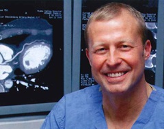
"Cardiac catheterization is still the gold standard for assessing narrowing of the coronary arteries, but 64-slice CT is a reliable tool for visualizing the presence of plaque without the risks associated with catheterization,” says Mukul Chandra, MD, cardiologist at MVH and associate clinical professor at Wright State University School of Medicine.
The new test may be especially useful for certain patients who have atypical chest discomfort and also have intermediate risk for heart disease. “Cardiologists are reluctant to take these patients to the cath lab, but nuclear studies and other non-invasive tests are often inconclusive,” says Dr. Chandra.
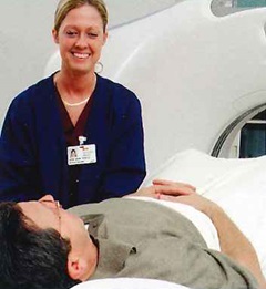
 The breakthrough of 64-slice CT is that it allows radiologists and medical imaging technologists to capture clear, stop-action images of the beating heart. Patients take medication before the testing to slow their heart rate slightly. As the images
are captured, contrast (dye) is injected through an IV placed in the arm. Patients also need to hold their breath for 15 to 20 seconds during the test.
Most patients are in and out of the hospital in 30 to 40 minutes. Processing the test data for interpretation takes several hours.
“CT technology has advanced very rapidly over the past several years,” says Larry Hall, DO, an MVH radiologist. “Combining this technology with the appropriate patient indications make this an important service to our medical staff and community.”

